If you have been offered embolisation for pelvic veins or a varicocele, you may have heard talk of pelvic embolization coils, foam or glue. These are the materials a specialist uses to close a faulty vein from the inside. This guide explains what each one is, how it works, why they are often combined, and what the evidence says about results and risks.
What does embolisation actually do?
In pelvic congestion, blood flows the wrong way down widened ovarian or pelvic veins and pools in the pelvis. In a varicocele, the same thing happens in the testicular vein. Embolisation blocks the faulty vein so that blood takes other, healthier routes back to the heart.
The specialist reaches the vein through a thin tube (catheter) passed in from the neck, groin or arm, guided by X-rays. A dye injection (venogram) shows which veins are refluxing. The chosen material is then placed through the catheter. You can read the step-by-step procedure in our guides to ovarian vein embolisation and varicocele embolisation.
Coils
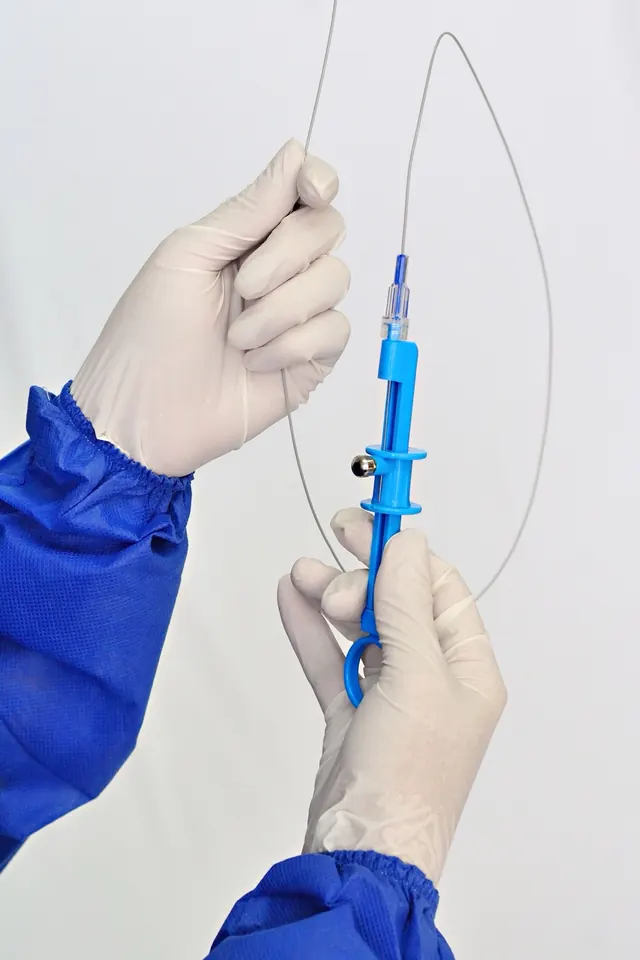
What they are. Tiny springs, usually of platinum or a similar metal, often with fine fibres attached. They are pushed out of the catheter in a straight line and curl up inside the vein.
How they work. The coil slows the blood, the fibres encourage a clot, and over the following weeks the vein scars shut around it.
When they are used. Coils are the most widely used material for the main ovarian vein and for internal iliac vein branches. Several are usually placed along a length of vein.
Downsides. Coils are the material most likely to move out of place (see below). A long run of coils can also be costly, and coils can cause a small blurred patch on some later scans of the pelvis.
Vascular plugs
What they are. Small, self-expanding cages of fine metal mesh, shaped like a cylinder or a series of discs.
How they work. The plug opens to fill the vein and blocks it in one spot. It is attached to a delivery wire until the specialist is happy with its position, so it can be pulled back and repositioned before release.
When they are used. Plugs suit wider veins, where a coil might not hold well. They are sometimes used with coils or foam behind them.
Sclerosant foam (sclerotherapy)
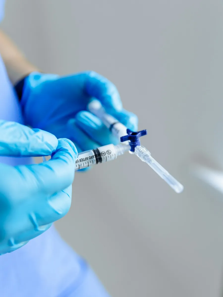
What it is. A medicine such as polidocanol or sodium tetradecyl sulfate (STS), mixed with air or gas to make a foam. The same medicines are used to treat leg and vulval varicose veins.
How it works. The foam pushes blood aside and irritates the inner lining of the vein. The vein becomes inflamed and then seals.
When it is used. Foam can flow into the tangle of small veins around the womb and ovaries that coils cannot reach. It is often injected before or between coils, a combination sometimes called a “sandwich”. Some teams use foam alone, and it is also used for vulval and thigh veins fed from the pelvis. See vulvar varicose veins treatment.
Downsides. A temporary ache after foam is common. Because it is a liquid, it must be controlled carefully so it does not travel where it is not wanted.
Medical glue
What it is. A liquid adhesive called a cyanoacrylate, the same family of chemicals used to close skin wounds.
How it works. It hardens within seconds of touching blood and seals the vein.
When it is used. Glue is used most often in varicocele, and in some centres for pelvic veins, alone or with coils. You may feel brief warmth or a cramp as it goes in.
Why materials are often combined
There is no single “right” material. The choice depends on:
- Vein size. Wide veins may need plugs or larger coils. Small branches suit foam.
- Vein pattern. Many women have several connected refluxing veins on both sides.
- The aim. Blocking the trunk alone can leave smaller side veins open. Adding foam treats those too.
- The team’s experience. Specialists tend to use the materials they know best.
A common approach is to close the main ovarian vein with coils or a plug and use foam for the smaller veins around it.
What does the evidence say?
The evidence for pelvic vein embolisation as a whole is of low quality. A 2016 systematic review by Daniels and colleagues found about 75% of women reported substantial early pain relief, but most studies were small case series without a comparison group. The European Society for Vascular Surgery’s 2022 guideline makes only a cautious recommendation (class IIb) to consider it in selected patients.
A larger 2026 review looked at 28 studies and 2,688 patients, grouped by material:
- Closing the vein worked in nearly every case with all materials (98.6% to 100%).
- Pain improved with every material, with clinical success between about 68% and 100% across groups.
- Coils had the highest rate of moving out of place, at about 2.6%.
- Plugs had few complications and low recurrence.
- Foam alone had the lowest recurrence but the most repeat procedures.
The authors stressed that the studies were small and did not compare materials directly, so these differences should be read with caution. In other words, there is no proven “best” material for everyone.
Risks to know about
Most risks are the same whichever material is used: bruising, an ache in the pelvis or back for a few days, a reaction to the contrast dye, and a small radiation dose. Risks more linked to the materials include:
- Coil or plug migration. The device moves from where it was placed, sometimes towards the heart or lungs. Reviews put this at under 2% to about 2.6% for coils. In one series of 218 women, 2 had a coil travel to the lung: one had no symptoms and the other coil was retrieved. Most migrations are seen straight away and can be retrieved through the same catheter.
- Coil protrusion. Part of a coil can stick out into a neighbouring vein.
- Non-target embolisation. Foam or glue reaching a vein it was not meant for.
- Clots and phlebitis. A clot in nearby pelvic veins, or a tender, inflamed vein, more often after foam.
One UK leaflet advises avoiding straining exercise for about six weeks, partly to reduce the chance of coils moving. Advice differs between centres, so follow your own team’s plan. See recovery after embolisation for what to expect.
Do coils stay forever? MRI and other questions
Coils and plugs are designed to stay in place permanently. Foam and glue do their work and the treated vein shrinks into scar tissue.
MRI scans. Most coils and plugs used today are made of metals considered suitable for MRI under set conditions. Even so:
- Tell the MRI team you have had embolisation.
- Ask your treating team for a record or implant card listing the devices used.
- Ask your specialist if you are unsure about a particular scan.
The metal can also blur the picture close to the coils on some scans, which is worth mentioning when a pelvic MRI is planned.
Questions to ask your specialist
- Which materials do you plan to use for me, and why?
- Will you treat one side or both, and the smaller veins too?
- What is your experience with coil migration, and how is it handled?
- Can I have a record of the devices for future scans?
For an overview of all the options, see pelvic congestion syndrome treatment and our guide to embolisation costs in Singapore. A Singapore vascular surgeon’s article on PCS treatment options also covers how embolisation fits in.
When to see a doctor
Contact your treating team if, after embolisation, you have pain that is getting worse rather than better, a fever above a low grade, redness or swelling at the entry site, or symptoms that return.
Get urgent medical help for sudden chest pain, breathlessness or coughing up blood, a swollen, painful or red leg, sudden severe pelvic or abdominal pain, or heavy bleeding from the entry site that does not stop with firm pressure. In Singapore, call 995 in an emergency.
Frequently asked questions
What are pelvic embolization coils made of?
They are small springs, usually made of platinum or a similar metal, and often coated with fine fibres that help a clot form. Once placed, they stay in the vein permanently and the body grows tissue around them.
Is ovarian vein embolization or sclerotherapy better?
They are usually not either-or. Many specialists use coils or plugs to block the main ovarian vein and a sclerosant foam to close the smaller veins around it. A 2026 review found that coils, plugs, foam and glue all closed veins in nearly every case, but the studies were small and did not compare the materials directly.
Is sclerotherapy for pelvic congestion syndrome done on its own?
Sometimes. Some teams use foam alone, especially for smaller veins or for vulval and leg veins fed from the pelvis. For a wide ovarian vein, foam is more often combined with coils or a plug so that it stays where it is needed.
Are ovarian vein embolisation coils safe in an MRI scanner?
Most coils and plugs in use today are made of metals that are considered suitable for MRI under set conditions. The exact product matters, so tell the MRI team you have had embolisation and, if you can, bring a record of the device names from your treating team.
What is glue embolization for varicocele?
It uses a liquid medical glue (a cyanoacrylate) that hardens within seconds of touching blood, sealing the testicular vein. It is often used in varicocele, alone or with coils. You may feel brief warmth or a cramp as it is injected.
How common is varicocele embolisation coil migration?
It is uncommon. Reviews of pelvic and testicular vein embolisation put coil migration at roughly 2 to 3 in 100 cases or fewer. Most are spotted straight away and retrieved, or cause no symptoms.
Sources and further reading
- Strobl F et al. Pelvic vein embolization using different embolic agents for pelvic venous insufficiency: a systematic review and meta-analysis. J Vasc Surg Venous Lymphat Disord 2026
- Daniels JP et al. Effectiveness of embolization or sclerotherapy of pelvic veins for reducing chronic pelvic pain: a systematic review. J Vasc Interv Radiol 2016
- BSIR — Pelvic congestion syndrome and pelvic vein embolisation (patient leaflet)
- Ratnam LA et al. Pelvic vein embolisation in the management of varicose veins. Cardiovasc Intervent Radiol 2008
- CIRSE — Pelvic congestion syndrome (patient information)
This article is general information, not personal medical advice. Please speak to a doctor about your own symptoms. How we create our content.