A pelvic venogram is an X-ray test of the veins in your pelvis. A specialist passes a thin tube into a vein, injects a contrast dye and watches on a screen how blood flows. It gives the most detailed picture of pelvic vein problems such as pelvic congestion syndrome, and it is usually done at the start of treatment rather than as a separate test. This guide explains what happens, why it is used, and how to prepare.
What is a pelvic venogram?
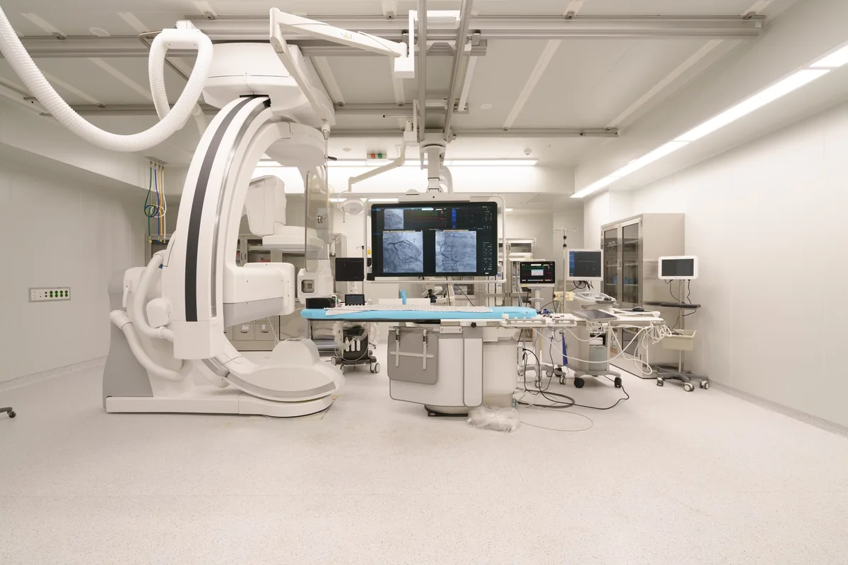
“Venogram” simply means a picture of the veins. In a catheter venogram (also called venography or phlebography), the dye goes directly into the veins being studied, through a fine, flexible tube called a catheter. Moving X-ray images (fluoroscopy) then show:
- Which veins are widened, and how much
- Whether blood is flowing backwards (reflux) down the ovarian or internal iliac veins
- Whether a vein higher up, such as the left kidney vein or the left iliac vein, is narrowed or squeezed
- Where the widened pelvic veins connect to veins in the vulva, buttocks or legs
Why is it called the reference standard?
Most specialist societies and reviews describe catheter venography as the reference standard, or “gold standard”, for pelvic venous disorders. There are three main reasons:
- It shows flow directly. Ultrasound, CT and MRI give strong clues, but a venogram shows dye actually running the wrong way down a vein.
- Positioning can be adjusted. The table can be tilted, or you may be asked to bear down, to make reflux easier to see.
- Pressures can be measured. If compression is suspected, the specialist can measure the pressure on each side of the narrowing.
There is one important catch. A venogram is invasive, so it is not used as a first test. Some specialists rely mainly on a careful duplex ultrasound and only use a venogram when treatment is planned. Our guide to how pelvic congestion syndrome is diagnosed explains the usual order of tests.
Where it fits with other tests
Usually you will have had other imaging first:
- Duplex ultrasound is safe, uses no radiation and can show reflux while you stand or strain. See our pelvic venous ultrasound guide.
- CT or MR venography maps the veins in 3D and checks the other organs. See CT and MR scans of the pelvic veins.
These scans help decide whether a venogram, and treatment, are worth doing. Widened veins are also common in women without symptoms, so imaging findings always need to fit your story.
Pelvic venogram with embolization or stenting
Because a venogram already involves a catheter inside the right veins, it makes sense to treat in the same sitting. If the pictures confirm refluxing veins that match your symptoms, the specialist can close them straight away with coils, plugs, foam or glue. This is ovarian vein embolisation.
If the main problem is a squeezed iliac vein, a stent may be placed to hold it open. Our guide to iliac vein stenting explains that procedure.
The plan should be agreed with you before the day. Ask what will happen if the venogram shows something unexpected, and whether treatment will go ahead in the same session or be discussed with you afterwards.
Pelvic venogram with IVUS
Intravascular ultrasound (IVUS) uses a tiny ultrasound probe on the end of a thin wire, passed inside the vein. It shows the vein from the inside, including its wall and true width. X-ray pictures can make a squeezed vein look normal when it is flattened from front to back, and IVUS helps avoid this.
IVUS is mainly used when May-Thurner syndrome or other iliac vein compression is suspected, and to choose the right stent size and position. It is not needed for every pelvic venogram.
What happens during the procedure
Details vary between centres, but a typical procedure goes like this:
- Getting ready. You change into a gown, and a small cannula is placed in your arm for fluids or sedation.
- Numbing the skin. Local anaesthetic is injected at the entry point, usually the groin, neck or arm. Many centres also give light sedation so you feel relaxed. A general anaesthetic is used in some.
- Placing the catheter. A small needle is placed in the vein, and a thin tube is guided up to the veins being studied under X-ray. You should not feel the tube moving inside you.
- Taking the pictures. Dye is injected into the kidney, ovarian and iliac veins in turn, and X-rays are taken. You may be asked to hold your breath or bear down.
- Treatment, if planned. Embolisation or stenting may follow.
- Finishing. The tube is removed and pressure is applied. Stitches are not usually needed.
A venogram alone may take under an hour. Combined with treatment, the whole procedure often takes one to two hours.
Contrast dye and radiation in plain words
The dye. The contrast dye contains iodine, which shows up on X-ray. When it is injected you may feel a warm flush or notice a metallic taste. These pass in seconds. Mild allergic reactions, such as itching or a rash, are uncommon, and serious reactions are rare. The team is trained to treat them. The dye is cleared by the kidneys, so your kidney function may be checked first.
Radiation. The X-rays involve a dose of radiation. The ovaries are close to the area being imaged, so teams keep the dose as low as they can, using short bursts of X-ray and shielding where possible. The small risk is weighed against the benefit of a clear diagnosis and treatment. You will have a pregnancy test beforehand if there is any chance you could be pregnant.
What are the risks?
A pelvic venogram is generally considered safe. Possible problems include:
- Bruising or a small swelling where the needle went in
- A reaction to the contrast dye
- Discomfort or a brief ache in the back or pelvis
- Rarely, infection, injury to the vein, or a blood clot
- If treatment is done at the same time, the risks of that treatment as well
Because the needle goes into a vein rather than an artery, serious bleeding is unusual.
How to prepare
Follow the instructions your own team gives you. In general:
- Tell the team about allergies (especially to contrast dye or iodine), kidney problems, diabetes and any chance of pregnancy.
- Medicines. Ask whether to pause blood thinners or diabetes medicines such as metformin. Do not stop anything without advice.
- Food and drink. You may be asked not to eat for a few hours if sedation is planned.
- Blood tests to check kidney function and clotting may be done.
- Getting home. Arrange for someone to take you home, as you should not drive after sedation.
- Questions. Write down what you want to ask, such as whether treatment will be done at the same time.
Afterwards
After a venogram alone, you rest for a short time while the entry point is checked, then usually go home the same day. Most people feel back to normal the next day. If embolisation was done too, read about recovery after pelvic vein embolisation.
Your specialist will explain what the pictures showed. Patient information from a Singapore vascular surgeon on PCS symptoms and diagnosis gives another overview of how these tests fit together.
When to see a doctor
See a doctor if you have pelvic pain that has lasted for months, pain that is worse after standing or during or after sex, or varicose veins around the vulva or upper thighs. Our pelvic symptom check can help you prepare for an appointment.
After a venogram, contact your team if the entry site becomes increasingly swollen, red or hot, keeps bleeding, or if you develop a fever. Get urgent medical help for heavy bleeding that does not stop with firm pressure, a swollen, painful leg, sudden breathlessness or chest pain, or signs of a severe allergic reaction such as swelling of the face or difficulty breathing. In Singapore, call 995 in an emergency.
Frequently asked questions
What is a pelvic venogram procedure like?
You lie on an X-ray table and a small area of skin in the groin, neck or arm is numbed. A thin tube is passed into the vein and guided to the pelvic veins, and dye is injected while X-ray pictures are taken. Most people feel pressure and a brief warm feeling from the dye, but little pain.
Is a pelvic venogram done with embolization at the same time?
Often, yes. If the venogram confirms refluxing veins that match your symptoms, and you have agreed to treatment beforehand, the specialist can close them with coils, plugs, foam or glue straight away. This avoids a second procedure. Some people have a diagnostic venogram only.
What is a pelvic venogram with IVUS?
IVUS is intravascular ultrasound: a tiny ultrasound probe on a thin wire is passed inside the vein. It shows the vein wall and measures how narrow the vein is more accurately than X-ray alone. It is most useful when compression of the iliac vein (May-Thurner syndrome) is suspected, and it helps size a stent.
What does ovarian vein radiology show?
Imaging of the ovarian veins looks at how wide they are, whether blood is flowing backwards (reflux) and whether there are widened veins around the womb and ovaries. Ultrasound, CT and MRI give indirect clues. A catheter venogram shows reflux directly when dye is injected into the vein.
How long does recovery from a pelvic venogram take?
After a venogram alone, most people rest for an hour or two and go home the same day, with normal activities the next day. If embolisation or stenting was done at the same time, recovery takes longer, and your team will give you specific advice.
Sources and further reading
- RadiologyInfo.org (RSNA/ACR) — Venography
- RadiologyInfo.org (RSNA/ACR) — Contrast materials
- Society of Interventional Radiology — Pelvic venous disorders (patient information)
- CIRSE — Pelvic congestion syndrome (patient information)
- Arnoldussen CW et al. Diagnostic imaging of pelvic congestive syndrome. Phlebology 2015
- Messina ML et al. Pelvic congestion syndrome as a differential diagnosis of chronic pelvic pain in women. Clinics (São Paulo) 2024
This article is general information, not personal medical advice. Please speak to a doctor about your own symptoms. How we create our content.