A pelvic congestion syndrome ultrasound is a scan of the pelvic veins and the way blood flows through them. It is usually the first test when doctors suspect that widened, leaky pelvic veins are behind long-term pelvic pain. It is not quite the same as the routine pelvic ultrasound many women have already had. This guide explains what is different, how to prepare, what the scan measures and what it cannot tell you.
Why a routine pelvic ultrasound can miss pelvic congestion
A standard pelvic ultrasound or transvaginal ultrasound is set up to look at the womb, the lining of the womb and the ovaries. It is very good for finding fibroids, cysts and many other causes of pain. But it is usually:
- Done lying flat. In this position the pelvic veins drain and can look normal.
- Focused on organs, not blood flow. The sonographer may not switch on the flow settings or follow the veins upwards.
- Short. Measuring veins and testing for reflux takes extra time.
So a “normal” routine scan does not rule out pelvic congestion. It may still be useful, because it can rule out other causes.
What is a pelvic venous duplex scan?
“Duplex” means the scan does two things at once. It shows a picture of the veins and measures blood flow with Doppler, often shown in colour, so the sonographer can see which way the blood is moving. A pelvic venous Doppler scan is the same idea.
For pelvic veins, the scan is usually done in two parts:
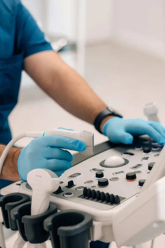
- Through the vagina (transvaginal). A slim probe gives close-up views of the veins around the womb and ovaries, and of veins crossing the womb wall.
- Through the tummy (transabdominal). This lets the sonographer follow the ovarian veins up towards the kidneys and check the left kidney vein and the iliac veins for compression.
Some centres also scan the groin, vulva and legs to look for “escape points” where pelvic veins feed visible veins outside the pelvis. This is especially useful if you have vulval or leg varicose veins.
Why you may be asked to stand, tilt or bear down
Pelvic veins fill and stretch when you are upright, and empty when you lie down. To show the problem, the sonographer may:
- Scan you standing, or on a couch tilted head-up
- Ask you to bear down, as if straining on the toilet, while holding your breath. This is called the Valsalva manoeuvre. It raises pressure in the tummy and shows whether blood rushes backwards down a leaky vein.
- Repeat measurements in more than one position
If a scan was done only lying flat, it is reasonable to ask whether these steps were included.
How to prepare
Preparation varies between centres, so follow your appointment letter. In general:
- Bladder. The abdominal part is sometimes done with a comfortably full bladder. The transvaginal part is usually done with an empty one.
- Food. Some centres ask you not to eat for a few hours beforehand, so that bowel gas does not block the view of the deeper veins.
- Clothing. Wear something easy to remove from the waist down. You will be given a gown or cover.
- Timing. Some specialists prefer to scan away from your period. Ask when booking.
- Comfort. You can ask for a chaperone, ask to insert the probe yourself, or ask the sonographer to stop at any time. If an internal scan is not right for you, ask whether an abdominal-only scan is possible.
The scan usually takes longer than a routine pelvic ultrasound, because the veins are measured in more than one position. Ask the centre how long to allow. It does not use radiation or dye, and you can drive and go back to normal activities straight afterwards.
What the scan looks at
The sonographer or doctor will usually check:
- The ovarian veins. How wide they are and whether blood flows backwards (reflux) down them, especially on the left.
- The veins around the womb and ovaries. Whether they are widened and twisted, known as pelvic varices.
- Veins crossing the womb wall. Widened veins here, sometimes called arcuate veins, are a typical finding.
- Flow speed. Very slow flow in the pelvic veins suggests pooling.
- Compression higher up. Narrowing of the left kidney vein (nutcracker) or the left iliac vein (May-Thurner).
- The ovaries. Some women with pelvic congestion have ovaries with many small follicles.
What counts as abnormal, in plain words
There is no single agreed set of numbers, and cut-offs differ between centres. Findings often used to support pelvic congestion include:
- Pelvic veins wider than about 4 to 5 mm
- An ovarian vein wider than about 5 to 6 mm, which is the usual answer to “what is a normal ovarian vein diameter?”
- Reflux: blood flowing backwards down the ovarian vein, or reflux brought on by bearing down
- Slow flow, roughly 3 cm per second or less, in the pelvic veins
- Widened veins crossing the womb, connecting the two sides
In one early study, women with pelvic congestion had a left ovarian vein of about 8 mm on average, compared with about 5 mm in women without symptoms. But there was overlap between the groups, and some women without pain also had reflux. This is why the numbers are a guide, not a verdict.
How to read your report
Reports may use terms such as “dilated tortuous parauterine veins”, “left ovarian vein incompetence”, “reflux on Valsalva” or “pelvic varices”. Taken together, these support a vein cause for your pain. A report may also say the veins are prominent but with no reflux, which is less likely to be significant.
Ask your doctor:
- Was the scan done standing, tilted or with bearing down?
- Which veins showed reflux, and on which side?
- Was there any compression of the kidney or iliac veins?
- Do these findings fit my symptoms?
Limits of the scan
Ultrasound is safe and cheap, but it has limits:
- It depends on the operator. Results vary with the experience of the person scanning. Reported accuracy varies widely between studies.
- Deep veins can be hard to see, especially with bowel gas or a higher body weight.
- It cannot prove the veins cause your pain. Widened veins and reflux are found in some women who feel well.
If the picture is unclear, or treatment is being planned, CT or MR venography may follow. A catheter venogram gives the most detail and is usually done at the time of treatment. Our pelvic congestion syndrome diagnosis guide covers these tests and the other causes doctors rule out, and our page on pelvic venous disorders explains how reflux and compression connect. A Singapore vascular surgeon’s PCS FAQs also cover what to expect from tests.
When to see a doctor
See a doctor if you have had pelvic pain for months, especially if it is worse after standing, during or after sex, or around periods. See the full list of pelvic congestion syndrome symptoms, or use our pelvic symptom check to organise your thoughts before an appointment.
Get urgent medical help for sudden severe pelvic or abdominal pain, heavy vaginal bleeding, fever with pelvic pain, fainting, or pain or bleeding when you might be pregnant. In Singapore, call 995 in an emergency.
Frequently asked questions
Can a pelvic ultrasound detect pelvic congestion syndrome?
A dedicated pelvic venous duplex scan can show the widened veins and backward flow that support the diagnosis. A routine pelvic ultrasound, done lying down to look at the womb and ovaries, often misses it. Ask whether your scan will look at the veins specifically.
What is a normal ovarian vein diameter?
There is no single agreed figure. Many specialists regard an ovarian vein wider than about 5 to 6 mm, especially with reflux, as abnormal. Diameter depends on your position and on how the vein is measured, so reflux and symptoms matter as much as size.
Is a transvaginal ultrasound for pelvic veins painful?
Usually not. The probe is about as wide as two fingers and is covered and lubricated. You may feel pressure, and some women with pelvic pain find it uncomfortable. You can ask for a chaperone, ask the sonographer to stop at any time, or ask whether an abdominal-only scan is possible.
What is a pelvic venous Doppler?
Doppler is the part of the ultrasound that shows blood flow and its direction, often in colour. A pelvic venous Doppler or duplex scan combines pictures of the veins with these flow measurements, so it can show whether blood is flowing the wrong way.
Do I need a full bladder for a pelvic vein scan?
It depends on the centre. The abdominal part is sometimes done with a comfortably full bladder, while the transvaginal part is usually done with an empty bladder. Follow the instructions in your appointment letter.
Sources and further reading
- Park SJ et al. Diagnosis of pelvic congestion syndrome using transabdominal and transvaginal sonography. AJR Am J Roentgenol 2004;182(3):683-8
- Barros FS et al. A comprehensive ultrasound approach to lower limb varicose veins and abdominal-pelvic connections. J Vasc Surg Venous Lymphat Disord 2024
- StatPearls — Pelvic Congestion Syndrome (NCBI Bookshelf)
- CIRSE — Pelvic congestion syndrome (patient information)
- Messina ML et al. Pelvic congestion syndrome as a differential diagnosis of chronic pelvic pain in women. Clinics (Sao Paulo) 2024
This article is general information, not personal medical advice. Please speak to a doctor about your own symptoms. How we create our content.