If an ultrasound has not given a clear answer, or treatment is being planned, your doctor may suggest a pelvic venous congestion CT or an MRI scan of the veins. These scans are called CT venography and MR venography. This guide explains what they show, what they miss, how to prepare, and where they fit alongside ultrasound and a catheter venogram.
What is venography?
“Venography” means a picture of the veins. For CT or MR venography, a contrast dye is usually injected into a vein in your arm, and the scan is timed so that the dye has reached the veins of the tummy and pelvis. The computer then builds a detailed 3D map.
- CT venography uses X-rays and an iodine-based dye. The scan itself takes only a few minutes.
- MR venography uses a strong magnet and radio waves, not X-rays. Some techniques use a gadolinium-based dye and some need no dye at all.
What CT and MR venography show
These scans can show:
- Widened ovarian veins, especially the left, and how wide they are along their length
- Pelvic varices: widened, twisted veins around the womb and ovaries
- Compression further up, such as the left kidney vein being squeezed (nutcracker) or the left iliac vein being squeezed (May-Thurner)
- Escape routes, where pelvic veins connect to veins on the vulva, buttocks or thighs
- The other organs: the womb, ovaries, kidneys, bladder and bowel
That last point matters. Pelvic pain has many causes, and a cross-sectional scan can show fibroids, ovarian cysts, signs of adenomyosis and other problems at the same time. Our guide to pelvic congestion syndrome diagnosis explains how doctors rule these out.
The big limitation: lying flat
Pelvic veins fill when you stand and drain when you lie down. CT and MRI are done lying on your back, so:
- Veins can look smaller than they really are during the day.
- A routine scan done for another reason may miss widened veins or mention them only briefly.
- Both scans give a snapshot, so they cannot reliably show which way blood is flowing. Reflux is better seen on duplex ultrasound or a catheter venogram.
Some MR techniques capture the dye moving through the veins over time, which can hint at reflux, but this is not available everywhere. A specialist will read the scan alongside your symptoms and other tests.
CT vs MRI: how they compare
| CT venography | MR venography | |
|---|---|---|
| How it works | X-rays | Magnet and radio waves |
| Radiation | Yes, a small dose | None |
| Dye | Iodine-based, through a drip | Gadolinium-based, or sometimes none |
| Time in scanner | A few minutes | Usually 15 minutes to an hour |
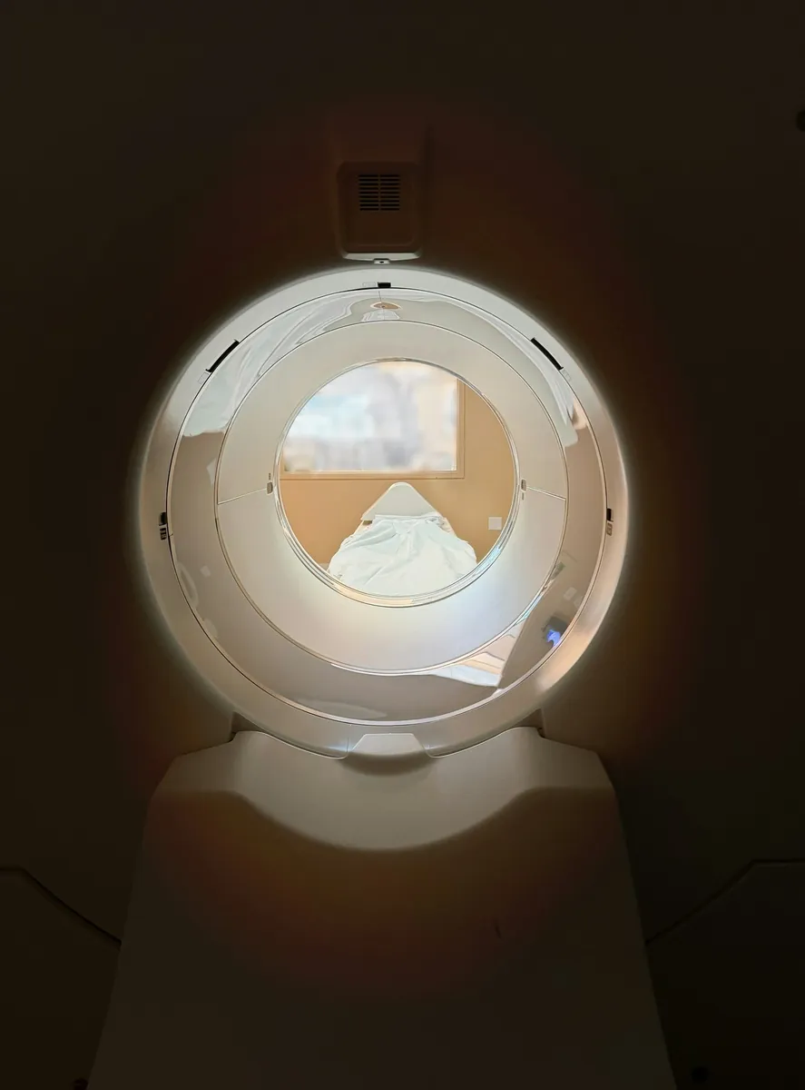
| Comfort | Open ring, quiet | Narrow tunnel, loud knocking noises |
| Metal implants | Usually no problem | Must be checked first |
| Availability | Widely available | Fewer slots, often costs more |
Pelvic congestion syndrome MRI is often described as the best non-invasive way to look at the pelvic veins, because it avoids radiation and shows soft tissue well. CT is quicker and easier to arrange, and is good at showing compression of the kidney and iliac veins.
Contrast dye and radiation
Contrast dye. When the dye goes in, you may feel warm, notice a metallic taste or feel as if you need to pass urine. These pass quickly. Allergic reactions are uncommon, and serious ones are rare. You may be asked to wait for a short time afterwards. Tell the team if you have had a reaction to dye before, or if you have kidney problems, as dye can affect kidney function.
Radiation. CT uses X-rays, which carry a very small chance of causing cancer later in life. Doctors weigh this against the benefit of the scan, especially for younger women and those who have many scans. MRI uses no radiation.
Pregnancy and breastfeeding. Always tell the team if you are or might be pregnant, or if you are breastfeeding.
How to prepare
Follow your appointment letter, as instructions vary. In general:
- Food and drink. You may be asked not to eat for a few hours beforehand.
- Blood test. Some centres check kidney function before giving dye.
- Metal. Remove jewellery and anything with metal. For MRI you will fill in a safety form about implants such as pacemakers, clips or embolisation coils.
- Feeling closed in. If you find enclosed spaces hard, tell the team before an MRI. They can talk you through it, and medicine to help you relax may be offered.
- Previous scans. Bring earlier reports or ask for them to be shared, so results can be compared.
Nutcracker and May-Thurner on CT or MRI
Patients often come across radiology articles on these conditions. In plain words:
- Nutcracker syndrome. The scan may show the left kidney vein narrowed where it passes between the aorta and another large artery, with a widened section before the narrowing and sometimes a widened left ovarian vein. Read more about nutcracker syndrome.
- May-Thurner syndrome. The scan may show the left common iliac vein flattened by the right iliac artery crossing in front of it, sometimes with extra collateral veins or old clot. Read more about May-Thurner syndrome.
Some narrowing in both places is common in people without symptoms, so a scan finding is not a diagnosis on its own. A Singapore vascular surgeon’s May-Thurner page explains how CT, MRI and venography are used together for iliac vein compression.
When CT or MR venography is used
The usual order of tests is:
- Duplex ultrasound first. It is safe, uses no radiation and can show reflux when you stand or bear down. See our guide to the pelvic congestion syndrome ultrasound.
- CT or MR venography when the ultrasound is unclear, when compression further up is suspected, when the veins need mapping before treatment, or when other organs need to be checked.
- A catheter venogram gives the most detailed picture and shows flow directly. It is invasive, so it is usually done at the start of treatment. See the pelvic venogram.
Sometimes the order is different. Widened pelvic veins are often first noticed on a CT done for another reason.
Questions to ask about your result
- Were the ovarian and pelvic veins widened, and on which side?
- Was there any compression of the kidney or iliac veins?
- Did the scan show anything else that could explain my pain?
- Do these findings fit my symptoms, and what is the next step?
Widened veins are found in some women without pain, so imaging is only one part of the picture. Our page on pelvic venous disorders explains how reflux and compression fit together.
When to see a doctor
See a doctor if you have pelvic pain that has lasted for months, pain that is worse after standing or during or after sex, or blood in your urine. You can use our pelvic symptom check to organise your thoughts before an appointment.
Get urgent medical help for sudden severe pelvic, abdominal or flank pain, a swollen, painful leg, sudden breathlessness or chest pain, heavy bleeding, or signs of a severe allergic reaction after a scan, such as swelling of the face or difficulty breathing. In Singapore, call 995 in an emergency.
Frequently asked questions
Can a CT scan show pelvic congestion syndrome?
A CT scan with contrast timed for the veins can show widened ovarian and pelvic veins and compression of the kidney or iliac veins. But it is done lying flat and gives a still picture, so it can underestimate the veins and cannot show reflux directly. A normal CT does not rule out pelvic congestion.
What does an ovarian vein CT show?
It shows how wide the ovarian veins are along their length, and whether there are widened veins around the womb and ovaries. A left ovarian vein that is clearly wider than usual and filled with dye early can suggest reflux, but the finding must fit your symptoms.
Is MRI better than CT for pelvic congestion?
MR venography is often seen as the best non-invasive test because it avoids radiation and gives good soft-tissue detail. CT is quicker and more widely available. Which one is used depends on the question being asked, local expertise and your situation.
What are pelvic varices on CT?
Pelvic varices are widened, twisted veins in the pelvis. On a CT report they may be described as dilated parauterine or periovarian veins. They are common, including in women without symptoms, so the report needs to be read alongside your symptoms.
How are nutcracker and May-Thurner syndromes seen on radiology?
CT or MR venography can show the left kidney vein squeezed between two arteries (nutcracker) or the left iliac vein squeezed by the artery crossing over it (May-Thurner). Because some narrowing is common in people without symptoms, the finding needs to match symptoms and is often confirmed with a venogram.
Sources and further reading
- Arnoldussen CW et al. Diagnostic imaging of pelvic congestive syndrome. Phlebology 2015
- Cochran RL et al. Body and extremity MR venography: technique, clinical applications, and advances. Magn Reson Imaging Clin N Am 2023
- CIRSE — Pelvic congestion syndrome (patient information)
- NHS — CT scan
- NHS — MRI scan
- Messina ML et al. Pelvic congestion syndrome as a differential diagnosis of chronic pelvic pain in women. Clinics (Sao Paulo) 2024
This article is general information, not personal medical advice. Please speak to a doctor about your own symptoms. How we create our content.