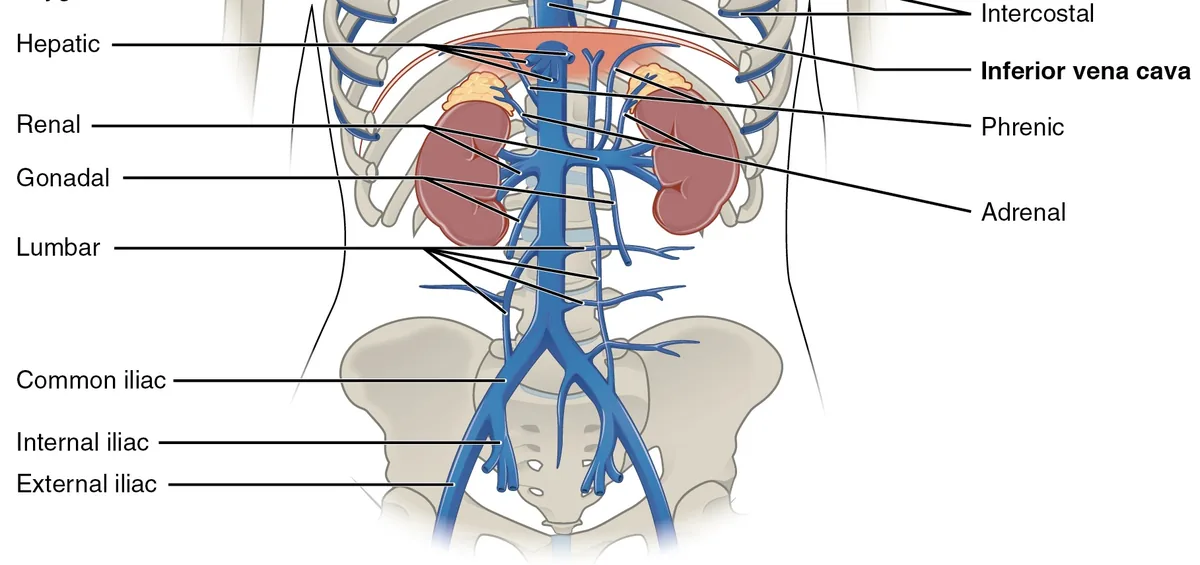
Understanding ovarian vein anatomy, and how the other pelvic veins connect, makes pelvic vein conditions much easier to follow. The pelvis drains through a small number of main veins, and the left side is built differently from the right. That difference explains why pelvic congestion, nutcracker and May-Thurner syndromes so often affect the left. This guide gives a simple, step-by-step tour, without the medical-school detail.
The big picture: two ways out
Blood from the womb, ovaries, bladder, bowel and pelvic floor collects in networks of thin-walled veins called plexuses. From there it has two main routes back to the heart:
- Upwards, through the two ovarian veins, which run up the back of the abdomen towards the kidneys.
- Sideways and back, through the two internal iliac veins, which join the leg veins to form the common iliac veins.
Both routes end in the inferior vena cava (IVC), the large vein running up the right side of the spine to the heart. Most pelvic blood normally leaves through the internal iliac route.
A simple map of the pelvic veins
Here is the layout as a list, starting at the pelvis and working upwards:
- Womb and ovaries
- Uterine and parametrial plexus (veins beside the womb) and ovarian plexus (veins beside each ovary)
- → Right ovarian vein → straight into the IVC
- → Left ovarian vein → into the left renal vein → crosses between the aorta and the superior mesenteric artery → IVC
- → Uterine veins → internal iliac veins (left and right)
- Pelvic floor, vulva, buttocks
- Internal pudendal, gluteal and obturator veins → internal iliac veins
- Legs
- Leg veins → external iliac veins in the groin
- Joining up
- Internal iliac + external iliac → common iliac vein on each side
- Left common iliac vein passes behind the right common iliac artery
- Left and right common iliac veins → IVC → heart
The ovarian veins: where each one drains
Each ovarian vein starts as a web of small veins beside the ovary, in the broad ligament that supports the womb. These merge into one or two channels that run upwards behind the bowel, alongside the ureter (the tube from the kidney to the bladder).
The two sides end differently:
- The right ovarian vein drains into the IVC directly, joining it at a gentle, slanting angle.
- The left ovarian vein drains into the left renal vein, joining it at close to a right angle. It is also a little longer than the right.
Small one-way valves, mostly in the lower part of each ovarian vein, normally stop blood falling back down. In a small minority of people the valves are missing, and this is more common on the left. “Gonadal vein” is simply the umbrella term: in men, the testicular veins follow the same pattern, which is why varicoceles are mostly left-sided.
The left renal vein
The left renal vein carries blood from the left kidney to the IVC. Because the IVC sits on the right, the left renal vein is longer than the right one and has to cross the midline. On the way it usually passes through a narrow gap between the aorta behind and the superior mesenteric artery in front.
As well as the left ovarian vein, it collects blood from the left adrenal gland and some veins of the back wall of the abdomen. If it is squeezed in that gap, pressure backs up into all of these branches, especially the left ovarian vein. When this causes symptoms, it is called nutcracker syndrome. In some people the vein runs behind the aorta instead, or splits around it, which can be squeezed in a different way.
The internal and common iliac veins
The internal iliac vein on each side collects blood from the pelvic organs, the pelvic floor, the vulva and perineum (via the internal pudendal veins), the buttocks (via the gluteal veins) and the inner thigh (via the obturator veins). These veins usually have few or no valves.
In the lower abdomen, the internal and external iliac veins join to form the common iliac vein. The two common iliac veins meet in front of the lower spine to form the IVC.
The left common iliac vein crosses behind the right common iliac artery, which can press it against the spine. When this narrowing causes symptoms, it is called May-Thurner syndrome. It can cause left leg swelling and clots, and it can also push blood back into the pelvic veins.
Escape points: how pelvic veins reach the vulva and thigh
The pelvic veins connect with veins outside the pelvis through a few small openings in the pelvic floor and groin. Specialists call them escape points or leak points. When pelvic veins are overfilled and refluxing, blood can leak out through them:
- Perineal (pudendal) point. The most common. Causes varicose veins on the vulva, perineum and upper inner thigh.
- Inguinal (round ligament) point. In the groin. Can cause veins in the groin, vulva and front of the thigh, and is sometimes mistaken for a hernia.
- Obturator point. Can feed veins on the inner thigh and perineum.
- Gluteal points. Can feed veins on the buttock and back of the thigh.
This is why some women have vulval varicose veins or unusual leg veins that come back after treatment. See leg varicose veins of pelvic origin.
Why the left side is more often affected
Put the pieces together and the left side has several extra weak spots:
- A longer left ovarian vein that meets the kidney vein at a near right angle
- Valves that are more often missing on the left
- A left renal vein that can be squeezed between two arteries
- A left common iliac vein that can be squeezed by an artery against the spine
Any of these can raise pressure in the left pelvic veins. That is why reflux in the left ovarian vein is the most common finding in pelvic congestion syndrome, although both sides and the internal iliac veins are often involved.
How this explains the symptoms
- A heavy, dragging ache that builds through the day. When you stand, gravity adds to the pressure in the pelvic veins, which have few working valves. Lying down lets them drain.
- Pain during or after sex, or around periods. The veins beside the womb and ovaries are stretched and swell further at these times.
- Visible veins on the vulva, buttock or thigh. Blood leaks out through the escape points.
- Blood in the urine with left flank pain. Points to the left renal vein being squeezed.
- Left leg swelling. Can point to the left common iliac vein.
For a full list, see pelvic congestion syndrome symptoms. A Singapore vascular surgeon’s overview of pelvic congestion syndrome also describes how these veins are involved.
When to see a doctor
Normal anatomy varies, and finding a wide vein on a scan does not always mean it is causing symptoms. See a doctor if you have a pelvic ache that has lasted months, pain that affects sex or daily life, new varicose veins on the vulva or upper thigh, or blood in your urine. Our pelvic symptom check can help you prepare.
Get urgent medical help for sudden severe pelvic or tummy pain, a suddenly swollen, painful leg, sudden breathlessness or chest pain, heavy bleeding, or fever with pelvic pain. In Singapore, call 995 in an emergency.
Frequently asked questions
What does the ovarian vein drain into?
It depends on the side. The right ovarian vein drains straight into the inferior vena cava, the body's main vein back to the heart. The left ovarian vein drains into the left renal vein, which then crosses in front of the aorta to reach the inferior vena cava.
What is the gonadal vein in women?
Gonadal vein is the general name for the vein that drains the sex glands. In women the gonadal veins are the ovarian veins; in men they are the testicular veins. Their layout is the same in both sexes, which is why men get left-sided varicoceles for the same reasons women get left-sided pelvic vein reflux.
Where is the iliac vein located?
The iliac veins lie deep in the lower abdomen and pelvis, behind the bowel and in front of the lower spine and pelvic bones. Each leg vein becomes the external iliac vein in the groin, joins the internal iliac vein from the pelvis, and forms the common iliac vein. The two common iliac veins join low in the abdomen, in front of the lower spine, to form the inferior vena cava.
Why is the left side more often affected?
The left ovarian vein is longer, joins the kidney vein at a near right angle and is more often missing valves. The left renal vein can be squeezed between the aorta and the superior mesenteric artery, and the left common iliac vein can be squeezed by the right iliac artery. All three push pressure back into the left pelvic veins.
Do the pelvic veins have valves?
The ovarian veins usually have a few one-way valves, mostly in their lower part, but a small proportion of people have none, more often on the left. The internal iliac veins usually have few or no valves. Missing or faulty valves let blood fall back into the pelvis.
Sources and further reading
- StatPearls — Anatomy, Abdomen and Pelvis, Renal Veins (PubMed record of the NCBI Bookshelf chapter)
- Jaworucka-Kaczorowska A, Simka M. Anatomical, pathophysiological and clinical aspects of extra-pelvic varicose veins of pelvic origin. Diagnostics 2025
- de Lorenzo Messina M et al. The left venous anatomy of the female pelvis and clinical outcomes: new insights. Rev Assoc Med Bras 2025
- Kurklinsky AK, Rooke TW. Nutcracker phenomenon and nutcracker syndrome. Mayo Clin Proc 2010
- Society of Interventional Radiology — Pelvic venous disorders (patient information)
This article is general information, not personal medical advice. Please speak to a doctor about your own symptoms. How we create our content.